-
 PIONEERING LEADERS.
CUTTING-EDGE TECHNOLOGY.
LIFE-CHANGING RESULTS.
PIONEERING LEADERS.
CUTTING-EDGE TECHNOLOGY.
LIFE-CHANGING RESULTS.
Welcome to Better Vision New Jersey
Welcome to Better Vision New Jersey, a dynamic, state-of-the-art ophthalmic practice that provides our patients with the highest quality of eye care possible. Our staff consists of some of the most experienced professionals in New Jersey, and our entire team is dedicated to improving and maintaining your eye health.
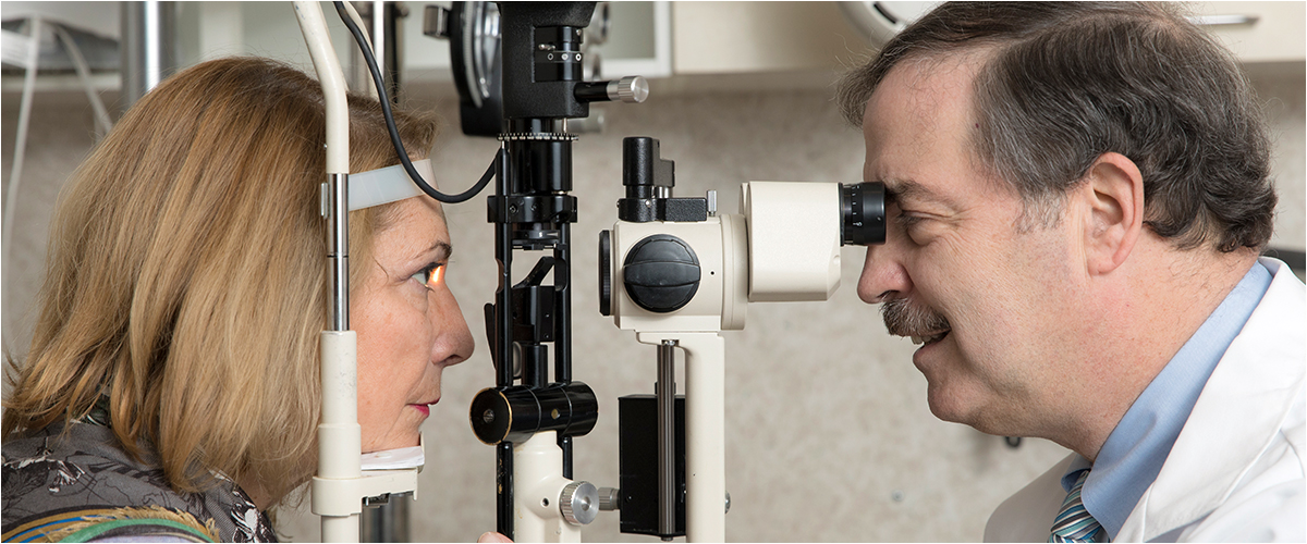
At Better Vision New Jersey, your health and experience with our practice are our only priorities.
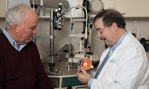
Since opening our Cranford, New Jersey location (formerly Cranford Ophthalmology) in 1987, we have consistently provided our patients with the best treatment options and results. We strive to best serve the needs of our New Jersey community and look forward to seeing you in your next visit our office. Our doctors—Dr. Joseph Calderone and Dr. Danica Yang—are fully dedicated to ensuring that our patients are satisfied with their results and have the visual assistance to live happier lives.
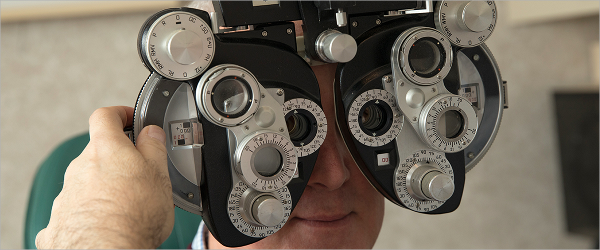
The eye care industry is constantly evolving, so we strive to use the latest and greatest available procedures and state-of-the-art technology. We are experts in all areas of eye care, but specialize in the treatment of dry eye and glaucoma, as well as cataracts. While we are able to effectively address all of these conditions, your general eye care is also deeply important to us. We strongly believe in the importance and value of optimal vision, and provide a variety of services to achieve this goal. Our doctors are dedicated to providing the best possible preventative care to help maintain your long-term eye health.
Welcome to Better Vision New Jersey
Welcome to Better Vision New Jersey, a dynamic, state-of-the-art ophthalmic practice that provides our patients with the highest quality of eye care possible. Our staff consists of some of the most experienced professionals in New Jersey, and our entire team is dedicated to improving and maintaining your eye health.
At Better Vision New Jersey, your health and experience with our practice are our only priorities.
Since opening our Cranford, New Jersey location (formerly Cranford Ophthalmology) in 1987, we have consistently provided our patients with the best treatment options and results. We strive to best serve the needs of our New Jersey community and look forward to seeing you in your next visit our office. Our doctors—Dr. Joseph Calderone and Dr. Danica Yang—are fully dedicated to ensuring that our patients are satisfied with their results and have the visual assistance to live happier lives.
The eye care industry is constantly evolving, so we strive to use the latest and greatest available procedures and state-of-the-art technology. We are experts in all areas of eye care, but specialize in the treatment of dry eye and glaucoma, as well as cataracts. While we are able to effectively address all of these conditions, your general eye care is also deeply important to us. We strongly believe in the importance and value of optimal vision, and provide a variety of services to achieve this goal. Our doctors are dedicated to providing the best possible preventative care to help maintain your long-term eye health.
30 YEARS OF EXCELLENCE IN EYE CARE
Cataracts
Are cataracts impairing your vision? Learn more about how our surgeons can clear up your vision with the latest in cataract treatment.

Dry Eye
Have red, itchy eyes? It may not be allergies. It might be Dry Eye Syndrome. Click to learn more about treatment options.
Glaucoma
Glaucoma is a sight-threatening disease that can cause serious damage to the optic nerve, which is a delicate, mesh-like structure that connects the eye to the brain.

OUR MISSION
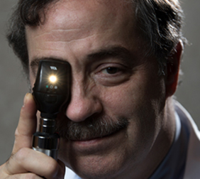
The physicians at Better Vision New Jersey are pioneering leaders in ophthalmology and are proud to provide the best possible eye care for patients of all ages. We are always excited to expand our family, and strive to build strong, lasting relationships with each and every one of our patients. Our mission is to provide world-class eye care in a patient-friendly, comfortable environment.
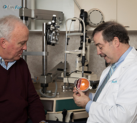
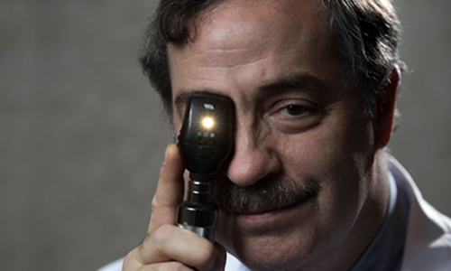
MEET OUR DOCTORS
For over three decades, the ophthalmologists at Better Vision New Jersey have dedicated their lives to improving the sight of thousands of patients. Our physicians are leaders in surgical innovation and are passionate about improving the lives of each and every patient.
What makes Better Vision New Jersey so exceptional? Read what our patients have to say.

I had cataracts and after Dr. Calderone operated on my eyes, my vision is now 20/20. The staff is extremely friendly and helpful. It’s a tremendously well-equipped practice in terms of the technology that’s used to take care of your eyes and the technical staff is also superb.
James O’Gorman
I have been a patient of Dr. Calderone’s for more than I can remember and I have always found the physicians and entire staff to be professional, caring and thorough in everything they do. All the physicians spend quality time with each patient to hear about their concerns or answer questions. I personally would highly recommend him to family and friends.
Sherry Villa
I had originally gone to the practice for floaters in my eye and discovered that I had cataracts. I found everyone at Better Vision New Jersey—from the physicians to the staff—to be extremely kind and caring. I was very impressed with the quality of care I received and how well they not only treated me, but also my husband. Their facilities are state-of-the-art and they are absolute experts in their field.
Mary Hannibal
CLICK ON OUR MAPS FOR DIRECTIONS TO OUR CRANFORD OFFICE
Better Vision Cranford
- 2 S. Ave East, Suite One
Cranford, NJ 07016.
Better Vision Cranford
- 2 S. Ave East, Suite One Cranford, NJ 07016.